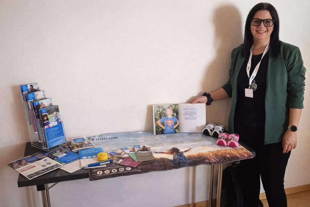
On March 6 and 7, 2026, the 16th Annual Conference of the Austrian Working Group for Interdisciplinary Treatment of Vascular Anomalies (AIVA) took place in St. Wolfgang am Wolfgangsee.
 A symposium held at one of Austria’s most beautiful locations. This naturally creates a blend of inspiration and focus—exactly what is needed to delve deeply into rare vascular diseases. Experts come together who otherwise rarely meet in such large numbers. Space is created for new perspectives—especially in the case of rare diseases, where every single case matters. The network is strengthened, which can later prove crucial in everyday clinical practice.
It is something special that we, as patient representatives for CMTC-OVM, are able to attend such a conference. It allows us to remind people that behind every diagnosis there is a person, not just a case report. Especially in the case of rare diseases, the exchange between medicine, research, and those affected is crucial.
Anatomical Basis of the Venous System and Its Disorders by Prof. Erich Brenner
The guiding structures in the deep venous system are the arteries, accompanied by nerves. The veins and lymphatic vessels form around them. In the deep system, the lymphatic vessels are directly connected to the arteries. Blood should flow from the inside outward in this system. If the venous valves are poorly developed or entirely absent, a backlog in blood flow occurs.
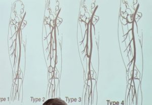
There are various anatomical variations: superficial vessels may be altered, leg veins may be completely absent, or they may be abnormally curved. How does this happen? Through a combination of embryology, genetics, and random developmental processes. Here, you can see various abnormalities of the venous system.
During embryonic development, the vascular system is not yet fully formed. Many vessels are temporarily created, which later fuse, regress, or are redirected. Vascular malformations occur when a vessel does not regress as usual or when another vessel persists abnormally. Genetic malformations can also cause a vessel to grow more strongly, branch differently, or form alternative pathways. In summary, vascular variations arise because the vascular system in the embryo is extremely flexible and complex, partly influenced by chance, and because developmental processes either fail to occur or do not regress as intended.
Dr. Alfred Obermayer about Venous Diagnostics
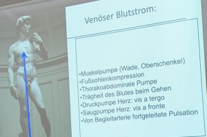
Venous problems are very common. Duplex ultrasonography is the most important examination in venous diagnostics. Blood cannot flow! It is not compressible. Using a muscle pump (raising and lowering the forefoot), the flow velocity in the vein can be measured accurately with duplex ultrasonography.
The cause of chronic venous insufficiency: we live on Earth in a force field. This force field exerts pressure on every mass. This pressure always flows downward—even in our bodies. Damage to the venous valves or permanently elevated venous pressure in the legs can prevent blood from returning sufficiently to the heart, causing it to accumulate in the leg veins.
Prim. Prof.Dr. René Müller-Wille On Classification and Diagnosis of Venous Malformations
He reports on congenital venous, arteriovenous, and lymphatic malformations. The diagnostic guidelines were finally published last year. This ISSVA classification forms the basis for the classification of vascular anomalies and is divided into two main groups: vascular tumors and vascular malformations (malformations of the vessels without tumor growth).

Benign congenital malformations of the venous system can develop with an abnormal network of vein-like vessels, thin vein walls, and reduced smooth muscle. The cause of venous malformation is genetic.
Symptoms include recurrent pain, thrombosis formation, and local complications such as fragility, bleeding in the intestinal area, or breathing difficulties if the malformation is located in the neck.
Visible symptoms include bluish spots, swelling, increased circumference, or dilated superficial veins. Interdisciplinary therapy is always particularly important for vascular malformations!
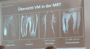 In the case of venous malformations, only the fluid can be visualized using the “T2-STIR” MRI examination. This is a special MRI scan in which fluids are displayed particularly clearly. In this sequence, the venous malformation can be seen very clearly, making it the best method for detecting slow-flow malformations.
Dr. Margitta Poetke about Laser Therapy for Venous Malformations
There is a wide spectrum of vascular malformations: capillary, venous, lymphatic, and mixed forms. It is important to classify the malformation correctly in order to apply the appropriate treatment. The focus is on reducing pain and functional limitations. The patient should feel comfortable in their own skin. This can be achieved through sclerotherapy, surgery, or laser therapy. The goal is to improve the condition, subjectively enhance the patient’s health, and, above all, slow the progression of the disease through laser therapy.
Different lasers are available for different treatment areas. Pulsed-dye laser treatments are used on the surface. For treatments under the skin, such as venous malformations, a laser with greater penetration depth is required. For example, a YAG laser can be used to selectively heat and close the vessels. The treatment is performed using an ice cube to effectively protect the skin. The ice cube must be moved to ensure that no hole forms.
“We cannot cure with the laser, but we can improve the situation for patients!”
Dr. Paolo Gasparella about the new ISSVA-classification
As previously described, the ISSVA classification forms the basis for the classification of vascular anomalies.
There was a need to revise the previous classifications. A working group was formed with five subgroups:
- Vascular tumors.
- Capillary malformations.
- Venous malformations.
- Lymphatic malformations.
- Arteriovenous malformations.

The ISSVA classification is a guideline for diagnosis, treatment, and further research. One thing is clear, however: a classification system is not set in stone. It must be continually updated and adapted.
It was a great conference, and we are grateful that we were able to attend again!